Rather than relying solely on black-box incubation experiments to quantify foraging, this research seeks an understanding of the underlying mechanisms, exploring how feeding currents are generated, and prey is detected, captured, and consumed or rejected across plankton types, from bacteria to larval fish.
While black box incubation experiments can be used to derive quantitative relationships between organisms, e.g., between predator and prey, they provide limited insights into the underlying mechanisms. One main activity is making direct observations of how organisms swim, navigate, forage, etc., and trying to understand the underlying mechanisms. Because plankton operate in ‘sticky’ water at low Reynolds number, formal physics is often required to interpret observations. The advent of high-speed video-microscopy has made it possible to record behaviors of all sorts at a relevant temporal and spatial scales, and cooperation with physicists has facilitated the understanding of underlying mechanisms. Physics models range from simple, idealized analytical models, to complex computational fluid dynamics modelling.

I wrote a book on the mechanistic approach, now partly outdated, but it had a nice cover illustration (Kiørboe (2008): A mechanistic approach to plankton ecology. Princeton University press).
One focus area has been foraging mechanisms in zooplankton and flagellates, and I have published two reviews that summarizes some of the work, a recent review on foraging mechanisms in phagotrophic flagellates and a not so recent review on zooplankton foraging mechanisms.

Ambush feeding copepods hang motionless in the water waiting for prey to pass by. Perceived prey are attacked. One would expect that the viscous boundary layer surrounding the copepod would push away the prey. That does not happen because the attack jump is faster than the time it takes for the viscous boundary layer to develop. The image shows the positions of the copepod and the prey immediately before (in white) and after (in black) the attack jump. The time difference between the two images is a few ms. From Kiørboe et al 2009

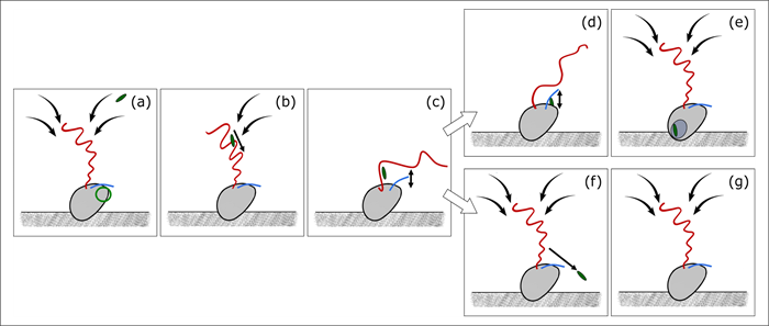
Cartoon based on high-speed video-microscopy illustrating how the flagellate Paraphysomonas foraminifera by the beating flagellum generates an incoming feeding current that brings in a bacterium that is captured (a-c) and either consumed (d-e) or rejected (f-g) after being handled (d,f). From Suzuki et al 2022